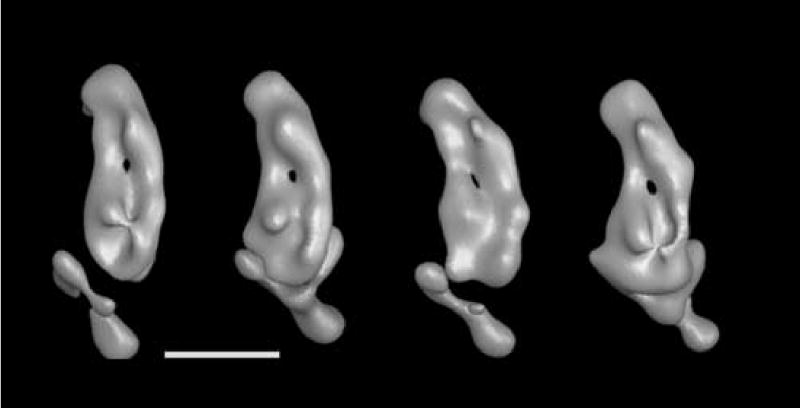
Figure 9.

From left to right: Volumes 35_2, 35_13, 35_15 and 35_20 from figure 8 shown here at a higher threshold value that reveals the groove in the cytoplasmic side of the membrane arm and the possible channel through the membrane arm. All views are from the cytoplasmic side of the membrane arm.
