
Fig. 2.
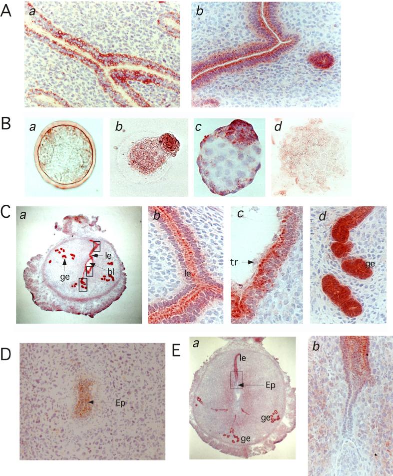
Immunohistochemistry of mouse uteri and embryos before, during and after implantation. A, mouse uterus from non-pregnant (a) and 4.5 day pregnant (b) mice. B, Pre-implantation stage mouse embryos. Morulae (a), blastocysts during hatching (b), a hatched and expanded blastocyst (c), and a hatched and one day cultured blastocyst (d). C, Day 4.5 pregnant mouse uterus with implanting blastocyst. Bystin was seen in the uterine luminal epithelial cells (le) and glandular epithelial cells (ge), at low (a) and high (b-d) magnification. Note that in luminal epithelial cells, bystin localizes to the apical side (b). In luminal epithelial cells adjacent to the blastocyst, bystin localizes to the apical side, whereas trophectoderm cells do not show bystin signals (c). Glandular epithelia cells strongly express bystin, whereas no polarization of bystin staining is seen in these cells (d). D, Implanted embryo at the epiblast stage or at day 5.5. E. Implanted embryo and uterus at day 6.5. ge, glandular epithelia; le, luminal epithelia; bl, blastocyst; tr, trophectoderm; ep, epiblast.