
Figure 2.
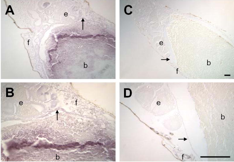
Nitrotyrosine (NTYR) levels in the midgut and surrounding tissues are increased in response to P. berghei infection. Immunohistochemical staining of 10μm sections of bloodfed A. stephensi was performed using polyclonal anti-nitrotyrosine (αNTYR). At 24h pBM, increased staining for NTYR (purple) was observed in tissues of P. berghei-infected (A, B) compared to uninfected A. stephensi (C, D). Staining was observed in the (b) blood mass, (e) ovarian tissue, (f) fat body and (→) midgut epithelium). The most pronounced staining occurred in blood meal bolus in the posterior midgut. Tissue samples were observed under 100x (A, C) and 400x (B, D) magnification; scale bar = 75μm.