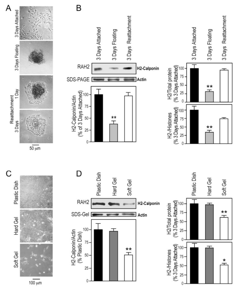
Figure 4.

Substrate anchorage- and stiffness-dependent expression of h2-calponin in alveolar cells. (A) SW1573 human alveolar cells were cultured on plastic dishes steadily or with vibration. Phase contrast microscopic images show that in contrast to the monolayer formed in the steady culture, continuous vibration prevented the cells from anchoring to the cultural dish and produced cell aggregates that could attach to the dish when vibration was stopped. (B) Total protein extracts from the cells were examined by SDS-PAGE and Western blotting using anti-h2-calponin polyclonal antibody RAH2. Normalized by the level of actin, total cellular protein, or histones (representing the number of cells), the results show that H2-calponin expression in alveolar cells decreased significantly when growing as floating aggregates in comparison with that in monolayer cells attached to plastic dish (*P<0.001). The expression of h2-calponin was up-regulated to resume the control level after the floating cells attached to the plastic substrate to form a monolayer. The results were summarized from 4 individual experiments. (C) SW1573 human alveolar cells were cultured on plastic dish or polyacrylamide gels of high or low stiffness for 3 days. (D) The levels of h2-calponin were determined by Western blot analysis using anti-h2-calponin antibody RAH2. Normalized by the amount of actin, total cellular protein, or histones, densitometry quantification shows a significant decrease in the expression of h2-calponin in alveolar cells grown on soft polyacrylamide gel in comparison with that in the hard gel and plastic dish cultures (*P<0.005, **P<0.001). The results were summarized from 3 individual experiments.
