
Fig. 1.
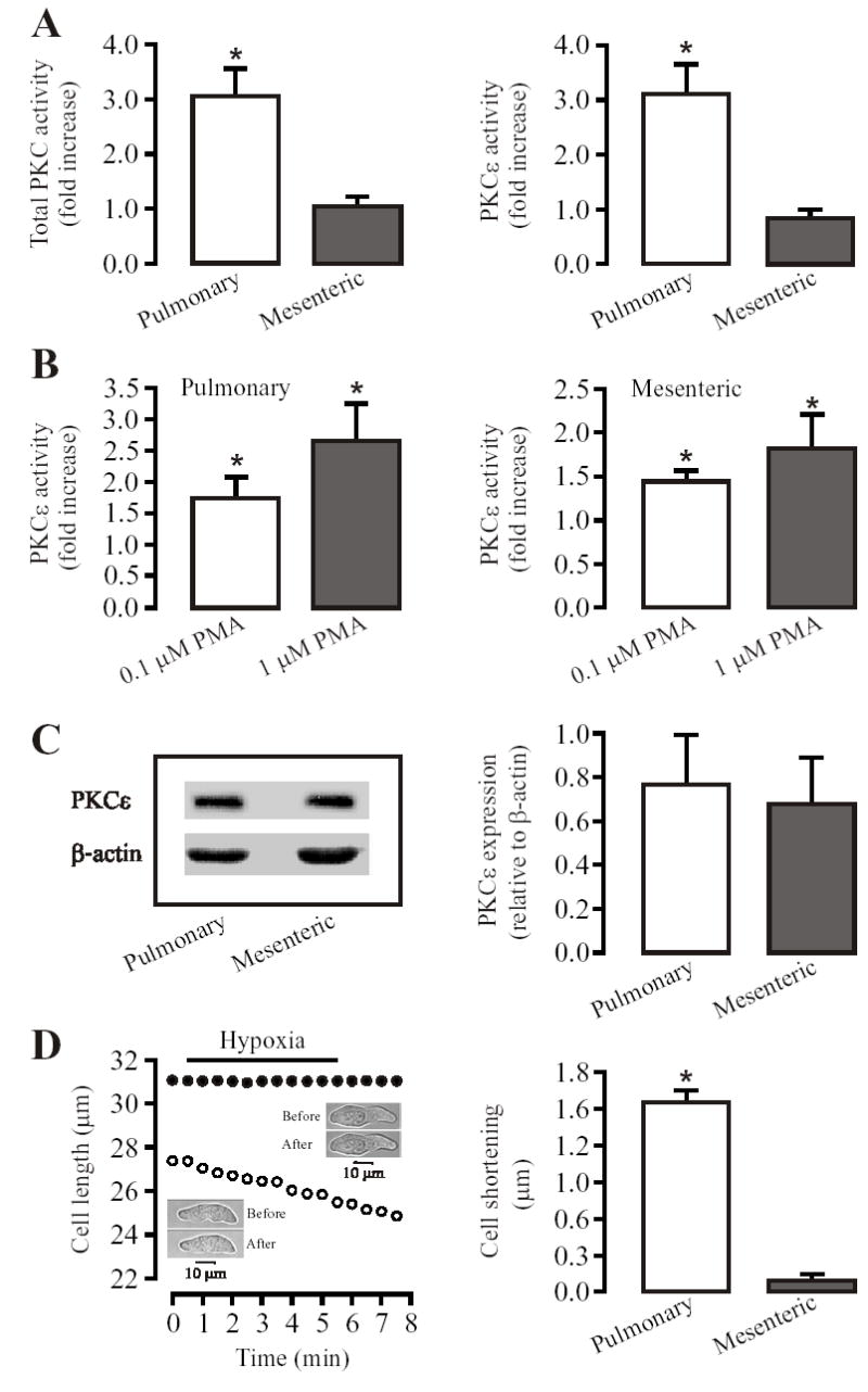
Acute hypoxia increases total PKC and PKCɛ activity in mouse resistance pulmonary, but not in mesenteric arteries. (A): Hypoxic exposure for 5 min caused an increase in the activity of total PKC and PKCɛ in pulmonary, but not mesenteric arteries. (B): The PKC activator PMA concentration-dependently augmented the activity of PKCɛ in both pulmonary and mesenteric arteries. (C): PKCɛ protein expression, determined by using SDS-PAGE and immunobloted with anti-PKCɛ antibody, was comparable in pulmonary and mesenteric arteries. Bar graph shows quantification of PKCɛ expression levels normalized to β-actin levels. All data are presented as means ± SE from 3 or 4 independent experiments. *P < 0.05 compared with control (without hypoxia or PMA). (D): Hypoxia for 5 min resulted in a significant contraction (shortening) in PASMCs, but not in MASMCs. Recording traces show that the effect of hypoxia on cell length in a PASMC (open circles) and MASMC (closed circles). Insets show that transmitted light images were taken before and after hypoxia for 5 min. Bar graph summarizes hypoxia-induced cell shortening in PASMCs and MASMCs. Data are presented as means ± SE from 21 PASMCs and 25 MASMCs in 4 independent experiments. *P < 0.05 compared with before hypoxia (normoxia).