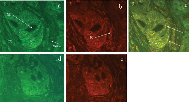
Figure 8.
Co-localisation of C4BP and CD40 in liver tumour tissue using dual immunofluorescence. Panels a–c shows a representative section of tumour tissue from a patient with cholangiocarcinoma stained for C4BP (green - FITC) and CD40 (red - PE ). In panel a, the arrows identify an epithelial ductular structure (DS) surrounded by tumour cells (TC) and stromal tissue. Positive C4BP staining is seen within the epithelia, tumour cells, and in mononuclear infiltrate in the surrounding stromal tissue. Panel b shows the same tissue section stained for CD40 with the arrow identifying the inflammatory cells within the surrounding stroma. Panels d and e shows a sequential section from the same specimen where the primary antibodies have been substituted for non immune serum (control). Panel c shows the merged image for panels a and b. The bright yellow areas indicate regions of C4BP and CD40 co-localisation within the epithelial cells of the ductular structure, many surrounding tumour cells, and the inflammatory cells within the surrounding stromal tissue.

