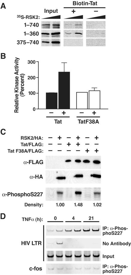
Figure 5. Activation of RSK2 by Tat.
(A) Autoradiography of radioactive in vitro synthesized RSK2 proteins before (Input) and after binding to biotinylated synthetic Tat (amino acids 1–72) or to beads alone. Increasing amounts of in vitro translated RSK2 were included in the binding reaction. (B) Kinase assay of endogenous RSK2 immunoprecipitated from Cos7 cells transfected with wild type Tat/FLAG, TatF38A/FLAG, or empty vector. Values are means±SEM of four experiments. (C) Western blotting of nuclear extracts isolated from Cos7 cells cotransfected with RSK2/HA and Tat/FLAG or with RSK2/HA and Tat F38A/FLAG constructs. Densitometric quantification of the phospho-S227-specific bands was performed using the NIH Image software. (D) Chromatin immunoprecipitation analysis of the Jurkat T cell line A2, latently infected with an HIV-based lentiviral vector expressing Tat/FLAG from the HIV LTR after treatment with TNF-α. At indicated time points, cells were harvested and immunoprecipitations were performed in duplicate with α-phospho-S227 antibodies followed by PCR with primers specific for the HIV LTR or the c-fos gene.

