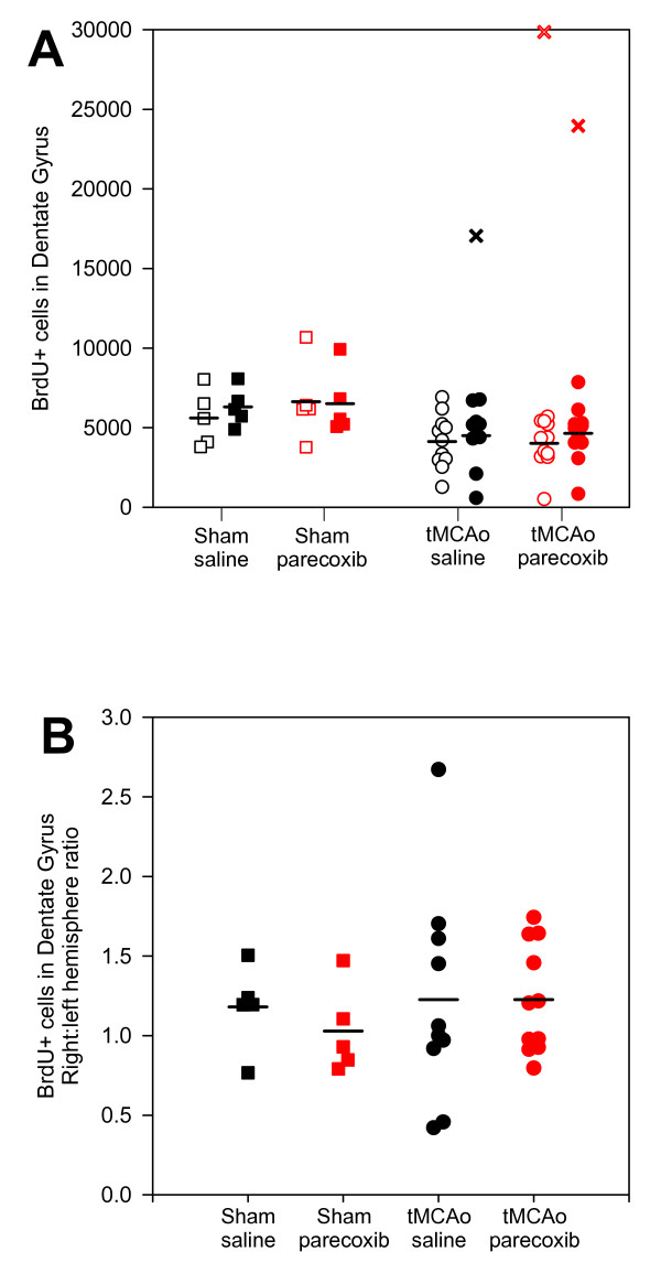
Figure 8.
BrdU incorporation in the dentate gyrus of the hippocampus. We estimated in average between four to six thousand BrdU-positive cells in the hippocampal DG (A). We revealed no significant differences between or within the four groups. The mean number of BrdU-positive cells was generally lower in the tMCAo groups than in the sham groups. However, if the DG was affected by ischemia (see Figure 9D to 9I) the BrdU incorporation increased dramatically (black × and red ×). Note that in one animal (marked with red ×) the ischemic injury also affected the contralateral hippocampus. B shows the BrdU incorporation ratio between the right and left hemispheres. The mean ratios for the four groups indicated no difference in the BrdU incorporation between the hemispheres or groups. Black □■: sham + saline; red □■: sham + parecoxib; black ○●: tMCAo + saline; and red ○●: tMCAo + parecoxib. The ischemic or sham (right) hemispheres are represented with filled symbols, whereas the contralateral (left) hemispheres are unfilled. Mean values are indicated with black horizontal bars.

