Figure 7.
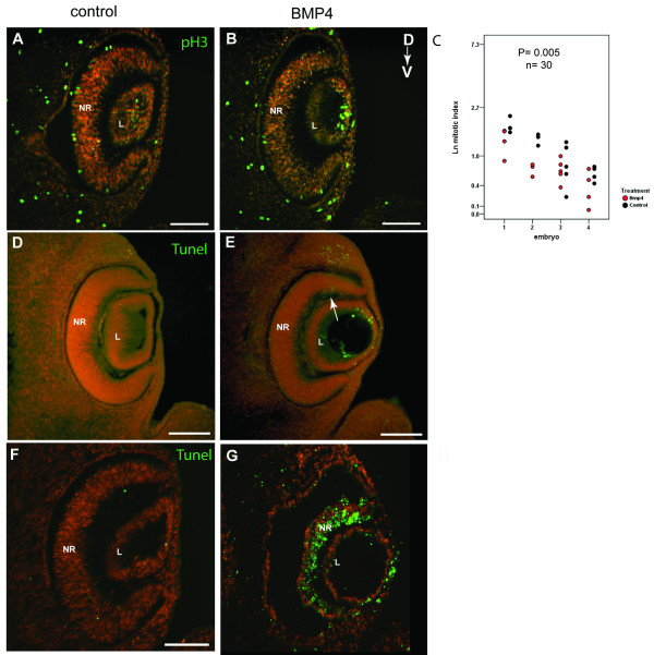
Effect of BMP4 treatment on proliferation and cell death. (A, B) pH3-positive cells (green) in retinal sections of control and BMP4-treated contralateral eyes of a post-culture embryo. Sections are counterstained with propidium iodide (red). (C) Graph showing the mitotic index per section per eye in BMP4-treated optic cups, compared with contralateral control optic cups (n = 30 sections from 4 embryos; p = 0.005 by ANCOVA). Data was Ln transformed for normalisation. Between embryo variability likely reflects the differences in growth rate of individual embryos. (D, E) Increased dorsal apoptosis detected by the TUNEL assay (green) in BMP4-treated eyes (arrow in E) compared to contralateral control eyes (D) of post-culture embryos. (F, G) Apoptosis detected in eyes of post-culture embryos after high dose BMP4 treatment. Widespread apoptosis detected throughout the neural retina. Scale bars: 0.1 mm. Abbreviations: D, dorsal; L, lens vesicle; NR, neural retina; pH3, phospho-histone H3; TUNEL, Terminal deoxynucleotidyl transferase-mediated dUTP Nick End Labeling; V, ventral.

