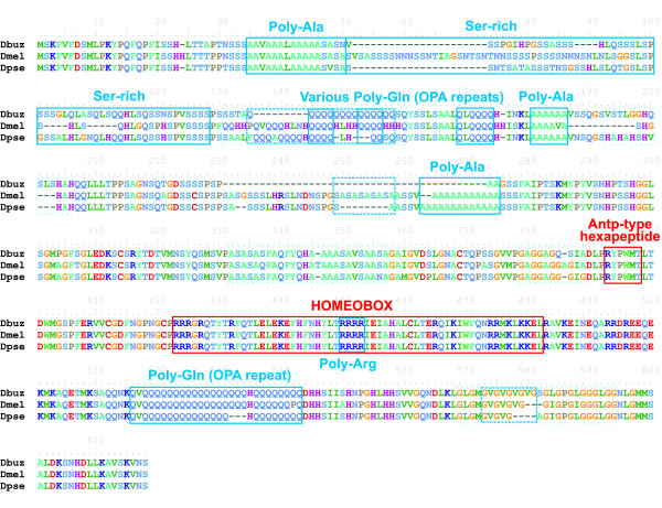
Figure 3.
Alignment of a Hox protein (ABD-A) showing multiple long repeats spacing functional domains. Functional domains are represented by red boxes, and repeats by blue boxes as follows: repetitive regions annotated in UniProt are represented by solid boxes, simple repeats by dashed boxes and complex repeats by dotted light boxes (see Methods). Notation: Dbuz = D. buzzatii; Dmel = D. melanogaster; Dpse = D. pseudoobscura.

