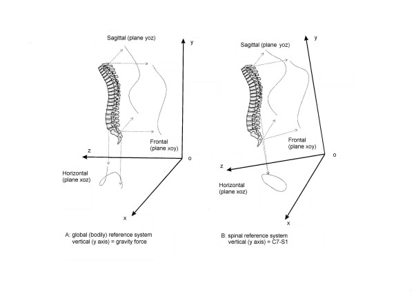
Figure 1.
3D representation of a real pathological spine (right thoracic, left lumbar scoliosis). In this figure the projections of the spine in the three spatial planes is reported: the frontal (xoy) plane is usually seen in the AP radiographs, the sagittal (yoz) is that of the classical LL x-rays, while the horizontal (yoz) plane (Top View) is not usually considered and it is the one studied here. The Top View doesn't allow to see the effect of the y axis, but joins together the sagittal and frontal plane deviations: in this respect it represents a useful auxiliary plane to have a quasi-3D projection of the spine. The Top View can be seen in a global (bodily) reference system (on the left: A) in which the vertical (y) axis is the gravity line, or in a spinal reference system (on the right: B) in which the vertical (y) axis is the line joining C7 and S1. In this last situation, that is the one that proved to be useful and it is adopted throughout this study, the entire reference system rotates with respect to the gravity line, as it can be seen on phthe right (B). These figures refer to the same single subject: note the differences between global (A) and spinal (B) Top Views.

