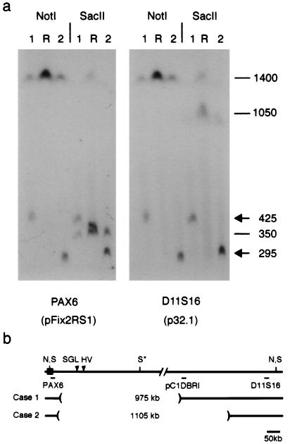
Figure 2.
Physical mapping of 3′ PAX6 deletions. (a) Pulsed-field gel of lymphoblastoid genomic DNA from cases 1 and 2 and a normal reference individual (R) hybridized with probes pFix2RS1 and p32.1. These probes detect one 1400-kb NotI (N) fragment and two SacII (S) fragments, which are 350 kb (centromeric) and 1050 kb (telomeric). The internal SacII (S*) site is partially cleaved (22, 32). The second SacII fragment in the reference (R) lane reflects a likely sequence polymorphism. (b) Restriction map showing hybridization probes and deletion breakpoints relative to PAX6. Probe pC1PBRI detects an altered NotI fragment in case 1 but not case 2 (data not shown). The deletions encompass breakpoints of the HV reciprocal translocation and the SGL paracentric inversion (arrowheads), which are located, respectively, 124 kb and 85–100 kb from the 3′ end of PAX6 (22, 33, 34).

