
Figure 3.
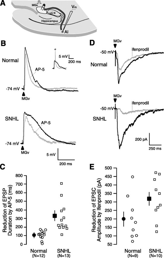
SNHL augments NMDA receptor function. A, A Schematic of the thalamocortical brain slice showing the position of a stimulating electrode in the MGv (square pulse), the pathway (dark line) from MGv to A1, and a recording electrode within A1 (Vm). The approximate distance the afferents travel from the MGv around the lateral geniculate (LGN) and hippocampus, radiating to the recording site in layer 2/3, is ∼5.5 mm. B, Maximum EPSP evoked by stimulating MGv (arrowhead). Note the significant duration (in milliseconds) reduction by the NMDA receptor antagonist AP-5 and a greater reduction in an SNHL neuron. Resting membrane potentials (in millivolts) indicated at the left of the traces. Inset shows MGv-evoked EPSP and EPSP-elicited spike (clipped); in all cases, the subthreshold maximum EPSPs were analyzed. C, Scatter plot of the magnitude of reduction in EPSP duration by AP-5 between normal and SNHL neurons shows a significantly greater AP-5-sensitive NMDA receptor-mediated component among SNHL cases. D, EPSCs evoked by stimulating at MGv (arrowhead). Note the reduction in amplitude by the NR2B subunit-specific antagonist ifenprodil and a greater reduction in an SNHL neuron. We do not rule out an effect of ifenprodil on presynaptic NMDA receptors. E, Scatter plot of the magnitude of reduction in EPSC amplitude by ifenprodil between normal and SNHL neurons shows a significantly greater ifenprodil-sensitive NR2B subunit component in SNHL neurons.