
Figure 1.
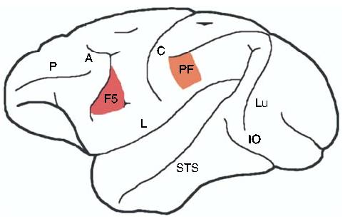
A schematic lateral view of the macaque monkey brain showing approximate regions of mirror neuron activity (adapted from [3]). Mirror neurons were first discovered in area F5 of the premotor cortex [1,2], and have also been reported in area PF of the inferior parietal lobule [23,24]. Labeled structures: A, arcuate sulcus; C, central sulcus; IO, inferior occipital sulcus; L, lateral fissure; Lu, lunate sulcus; P, principal sulcus; STS, superior temporal sulcus.