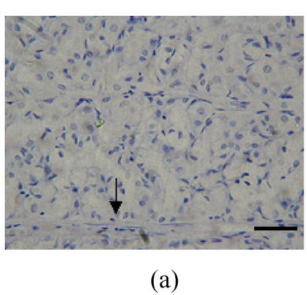
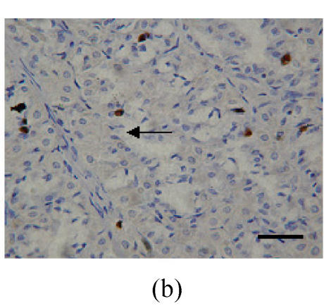
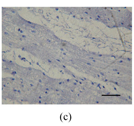
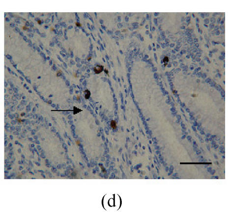
Fig. 1.
Immunohistochemical localization of FoxO proteins in the stomach of pigs (Bar=50 m). (a) Fundic gland control (×400). ↓: Muscularis mucosa; (b) Localization of FoxO4 in the fundic gland (×400). Localizations of FoxO4 in fundic part are very distinct, and primarily concentrated in the cell nuclei of the gastric mucosa. ←: Fundic gland; (c) Localization of FoxO4 in the non-glandular zones (×400). Compared with the non-glandular zones of control, FoxO4 staining was not observed; (d) Localization of FoxO4 in the pyloric gland (×400). FoxO4 in the pyloric gland was very similar to that in the fundic part, with the localizations concentrated in the cell nuclei of the mucosa. →: Pyloric gland


