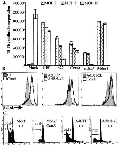
Figure 4.
Adenoviral-mediated manipulation of T cell proliferation and survival pathways. (A) The expression of p27KIP, CrmA, and IκB can block T cell proliferation. Lymphocytes from CAR Tg mice (BALB/c) were harvested and either mock transduced or transduced with adenovectors expressing either p27, CrmA, IκB, Mdm2, or GFP at the indicated MOI. One day after transduction, T cells were cultured with PMA and Con A in RP10 with [3H]thymidine for 2 days, and the amount of incorporated [3H]thymidine (±SE) was determined by scintillation counting. The first two bars represent mock-transduced cells that did not receive PMA or Con A. (B) Adenoviral-mediated expression of Bcl-xL in T cells. Lymphocytes from CARΔ1 Tg mice (BALB/c) were harvested and transduced with AdUbC-GFP or AdCMV-Bcl-xL at a MOI of 10, and then cultured in RP10 for 2 days. (Left) Cells were mock transduced and cultured without (−) or with 4 μg/ml Con A. Cells were stained with anti-B220, fixed, permeabilized, and then stained with an antibody to Bcl-xL. The cells were analyzed by flow cytometry for the expression of Bcl-xL in T cells (B220 negative). (C) Bcl-xL expression increases T cell survival in the absence of mitogenic stimulation. Apoptosis was determined in the cells from the same experiments described in B. The cells were harvested, stained with allophycocyanin-linked anti-B220, stained with propidium iodide, and DNA content (x axis) was determined in T cells (B220 negative) by flow cytometry. The percentage of T cells with a sub-G1 DNA content (apoptotic) representing the average of two experiments (±SE) is indicated.

