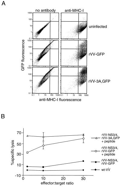
Figure 3.
(A) MHC I abundance on target-cell surfaces in the presence and absence of poliovirus 3A protein expression. Chimpanzee target cells were either uninfected or infected with rVV-GFP or rVV-3A,GFP at a multiplicity of infection less than 1 pfu per cell. At 12 h after infection, the cells were stained with anti-MHC I antibodies and analyzed by FACS. For each sample, 104 cells were plotted for red (MHC I) versus green (GFP) fluorescence. (B) Effect of exogenously loaded peptide on CTL-mediated lysis of 3A-expressing target cells. Chimpanzee target cells were infected with wild-type vaccinia (wt-VV) or coinfected with the viruses indicated in the presence or absence of the antigenic peptide GAVQNEITL as shown. Standard deviations of duplicated CTL assays are indicated when possible given the symbol size.

