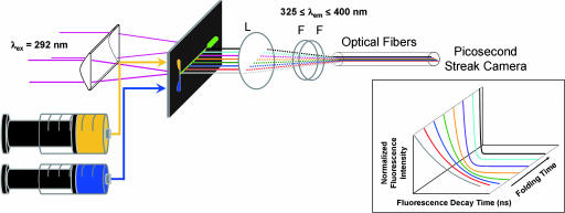
Fig. 5.
Schematic of the system used for FET kinetics measurements during protein folding. A 200-μm-thick stainless mixing plate is placed between two 3-mm-thick quartz windows (which are compressed by a stainless holder, not shown). Fluorescence from the flow channel is focused onto a bundle of optical fibers (≈0.1-mm diameter) and directed to a picosecond streak camera. L, lens; F, dielectric filter. (Inset) Image of the experimental data detected by a picosecond streak camera.

