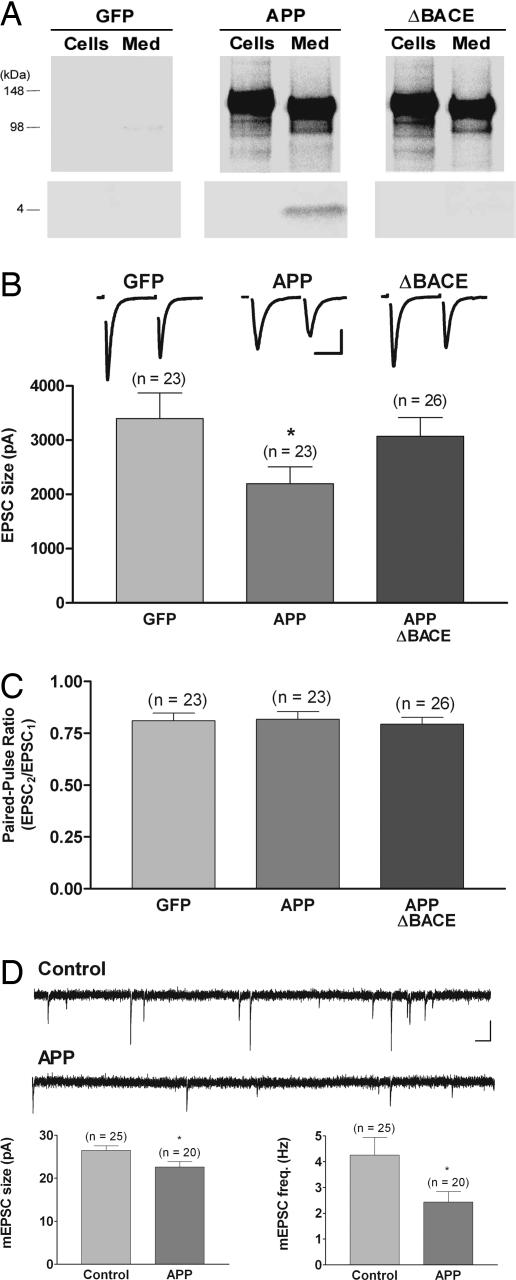
Fig. 1.
Overexpression of wild-type APP but not a mutant APP that is unable to produce Aβ reduces excitatory postsynaptic current (EPSC) size without altering the paired-pulse ratio (PPR); overexpression of APP also reduces mEPSC amplitude and frequency. (A) Immunoprecipitation by using 6E10 of [35S]methionine-labeled APP (Upper) and Aβ (Lower) from cells and medium showed that infection with viral constructs encoding either APP or APPΔBACE directed expression of full-length, cell-associated APP and secreted APPsα in medium. Viral constructs encoding GFP served as a control for these experiments. Only neurons overexpressing wild-type APP generated significant levels of Aβ. (B) Average EPSC size was reduced in neurons overexpressing wild-type APP (0.62 of GFP-alone control; ∗, P < 0.05) but not in neurons overexpressing APPΔBACE. Traces show typical paired-pulse responses from each group with action currents blanked for clarity. (Scale bar, 1.5 nA, 10 ms.) (C) There was no difference in the PPR, measured as the relative sizes of the first and second responses to a pair of stimuli delivered 50 ms apart, in neurons overexpressing wild-type APP or APPΔBACE compared with GFP-alone-expressing neurons. (D) There was a small but significant decrease in mEPSC size (0.85 of control; ∗, P < 0.02) and a reduction in mEPSC frequency (0.56 of control; ∗, P < 0.04) in neurons overexpressing wild-type APP. For these mEPSC experiments, results from uninfected and GFP-expressing neurons, which were indistinguishable, were combined for the control group. Traces show typical spontaneous responses from control and APP-overexpressing neurons. (Scale bar, 20 pA, 100 ms.)

