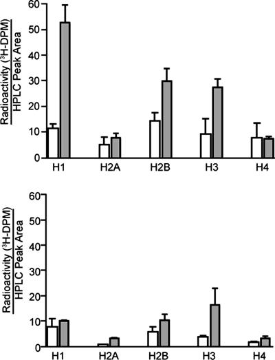
Fig. 3.
HPLC analysis of neocarzinostatin-induced 3H-labeling of histones from TK6 cells with DNA containing 5′-[3H]thymidine (Upper) or methyl-[3H]thymidine (Lower). Isolated nuclei were treated with neocarzinostatin (30 μM; shaded bars) or methanol vehicle (open bars), followed by histone extraction, nuclease digestion, and HPLC separation, as described in Materials and Methods. Tritium associated with the various histone-containing HPLC fractions was quantified by scintillation counting and reported as disintegrations per minute (DPM) per unit area of UV absorbance signal. Data represent mean ± SD for three independent experiments.

