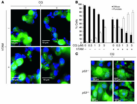
Figure 4. Effects of p53 activation with and without CQ on LC3 relocalization.
(A–C) GFP-LC3 fluorescence. Green, GFP-LC3; blue, DAPI. (A) A bulk population of primary Myc/p53ERTAM lymphoma cells with stable expression of the GFP-LC3 fusion protein was treated with and without 250 nM hTAM and with and without 5 μM CQ. Cell culture medium was changed daily. Cells were fixed and imaged using fluorescence microscopy at 24 and 48 hours. Representative images of cells at 48 hours are presented. (B) Quantification of the percentage of cells with more than 4 GFP-LC3 puncta per cell (punctate) compared with those with less than 4 GFP-LC3 puncta per cell (diffuse) treated with increasing doses of CQ with and without hTAM at 24 hours. (C) CQ modulates autophagy in a p53-independent manner. p53+/+ and p53–/– MEFs expressing GFP-LC3 were treated with CQ. Cells were fixed and imaged at 24 hours.

