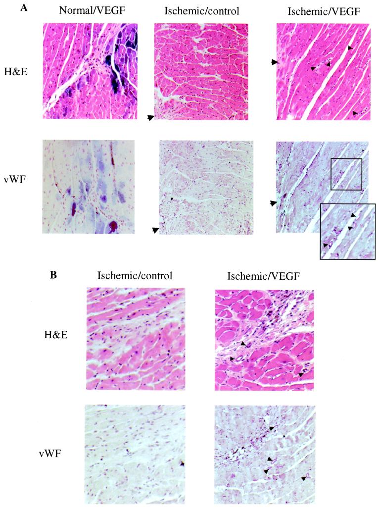
Figure 2.
Photomicrographs of normal and ischemic hearts inoculated with AAV-VEGF (Normal/VEGF and Ischemic/VEGF) and ischemic heart not injected with AAV vector (Ischemic/control). Arrows, infarcted regions; arrowheads, small blood vessels. (A) Area around the infarcted regions. (Upper) H&E staining. (Lower) vWF staining. An increase in vessel formation is observed in the Ischemic/VEGF hearts. An inset in the micrograph of the vWF-stained ischemic/VEGF heart presents an enlarged area to show blood vessels more clearly. (B) Area around scar tissues formed by needle injections. H&E and vWF stains of cardiac myocardium of both the Ischemic/control and Ischemic/VEGF mice. Note the new blood vessel formation around the scar caused by the needles in the AAV-VEGF-injected heart but not in the control without injection of AAV.

