
Fig. 4a, b.
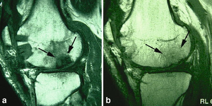
Sagittal T1-weighted SE images of the normal development of autologous osteochondral transplants. a Marked oedema in and around the osteochondral plugs at the recipient site 12 weeks (arrows) after surgery and b bony incorporation of the grafts with fatty bone marrow in and around the grafts (arrows) and filling of the donor site with cancellous bone after 2 years