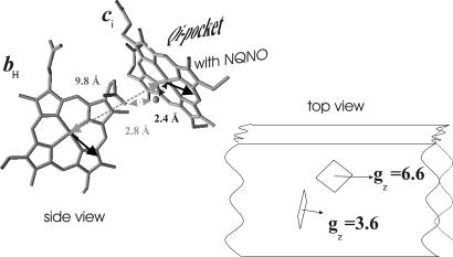
Fig. 7.
g-tensor orientation with respect to the 3D structure. (Left) Structure of the ci/bH pair. The oxygen atom bridging heme ci's central Fe atom to a propionate side-chain oxygen of heme bH (Protein Data Bank ID code ) is highlighted. Bold arrows indicate the orientations of bH's gz and ci's gx directions. (Right) Schematic representation of the mutual orientations of the ci/bH pair and their maximal g values as seen from the n-side of the membrane.

