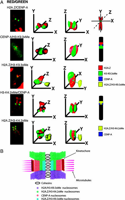
Fig. 3.
3D deconvolution analysis of the human inactive X chromosome centromere. (A) Human inactive X metaphase chromosomes in HEK 293 cells were immunostained with different pairwise combination of antibodies that recognize H2A.Z, CENP-A, trimethyl K9 H3, or dimethyl K4 H3, analyzed by 3D deconvolution microscopy and modeled to determine the spatial position of domains containing H2A.Z, CENP-A, trimethyl K9 H3, and dimethyl K4 H3. For each combination of antibodies shown, five to eight metaphase chromosomes were analyzed and modeled. (B) A model for the folding of pericentric and centric chromatin fibers into the unique 3D organization of the human inactive X chromosome centromere highlighting the spatial position of H2A.Z containing chromatin. This diagram has been modified from Sullivan and Karpen (3).

