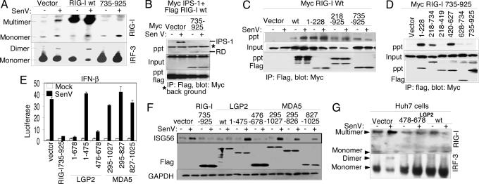
Fig. 3.
Mechanism of regulation by the RIG-I RD. Where indicated, cells were mock-infected or infected with SenV for 16 h before harvesting. (A) Stable Huh7 cell lines expressing vector alone, RIG-I wt, or RIG-I 735–925 were mock-infected or infected with SenV, and protein extracts were subjected to native PAGE and immunoblot analysis with anti-RIG-I antibody (Upper) or anti-IRF-3 antibody (Lower). Dimer/multimer and monomer protein forms are indicated. (B–D) Huh7 cells were cotransfected with plasmids encoding Myc-IPS-1, Flag-RIG-I wt, and vector or Myc-RIG-I 735–925 (B); Myc-RIG-I wt and the indicated Flag construct (C); or Myc-RIG-I 735–925 and the indicated Flag construct (D). Cells were infected as shown and harvested, and extracts were analyzed by immunoprecipitation (IP) and immunoblot assays. Shown are the abundance of Myc-tagged protein within anti-Flag IP products (Top), input Myc-tagged protein (Middle), and input Flag-tagged protein (Bottom). (E and F) Huh 7 cells were transfected with Renilla luciferase, IFN-β-luciferase plasmids, and plasmids encoding vector alone or the indicated Flag-tagged RIG-I, LGP2, or MDA5 constructs. After SenV infection, the cells were harvested and extracts were subjected to dual luciferase assay (E) (bars show relative luciferase and SD) and to immunoblot assay for abundance of ISG56, Flag-tagged protein (Flag), and GAPDH (F). (G) Anti-RIG-I (Upper) or anti-IRF-3 (Lower) immunoblot of Huh7 cell extracts separated by native PAGE. Protein monomer and multimer/dimer forms are indicated. Cells were transfected with vector control or expression plasmid encoding Flag-LGP2 478–378 or Flag-LGP2 wt. Extracts were prepared after SenV or mock infection.

