Fig. 1.
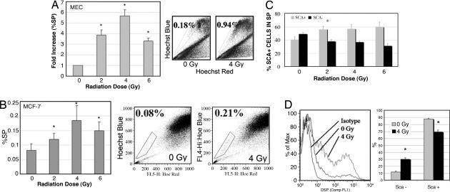
Clinically relevant doses of radiation increased the percentage of progenitor cells (%SP and Sca1+) in primary MEC culture and human MCF-7 cells. (A) MECs were isolated from BALB/c mice, cultured for 3 days, irradiated, and analyzed for %SP by Hoechst 33342 staining and flow cytometry. Radiation selectively increased the progenitor fraction (%SP) (P = 0.015 for 2 Gy, 0.008 for 4 Gy, and 0.05 for 6 Gy by the two-tailed t test). (B) MCF-7 cells were analyzed for %SP by Hoechst 33342 staining and flow cytometry. Radiation selectively increased the progenitor fraction (%SP) (P = 0.05 for 0 Gy vs. 4 Gy by the two-tailed t test). (C) Cells were analyzed for Sca1 in the SP 24 h after irradiation. Radiation selectively increased the Sca1+ (progenitor) fraction within the SP by killing the more sensitive Sca1− (nonprogenitor) cells (P < 0.05 for Sca1+ to Sca1− at 0 Gy vs. 2–8 Gy). The differences in effects of doses of 2 Gy vs. higher doses were not significant. (D) Anesthetized BALB/c mice were immobilized supine, and mammary glands (entire ventral surface) were irradiated. MECs were isolated 48 h after irradiation and analyzed immediately for Sca1 by flow cytometry. Radiation selectively increased the Sca1+ (progenitor) fraction and decreased the Sca1− (nonprogenitor) cells. ∗, P < 0.0001.

