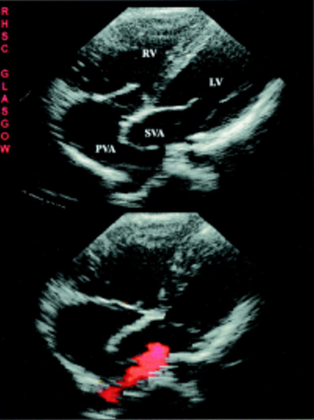
Figure 33 .

Parasternal four chamber views of patient with a Mustard procedure and pulmonary to systemic baffle leak. The upper image is in end diastole and shows the right ventricle (RV) to be enlarged and hypertrophied but the left ventricular (LV) size is not reduced because of a pulmonary to systemic shunt. A right sided pulmonary vein is seen draining into the pulmonary venous chamber. The lower frame is taken in systole and colour Doppler shows a low velocity shunt through the baffle leak.
