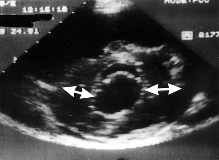
Figure 3 .
Parasternal short axis view of the left ventricle at the mitral valve level demonstrating a typical eccentric form of ventricular hypertrophy localised essentially at the lateral wall and posterior septum, while the anterior and posterior walls are normal. Reproduced from Nihoyannopoulos and McKenna with permission of Churchill Livingstone.10

