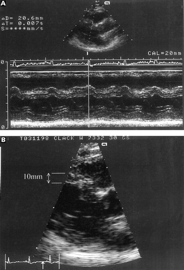
Figure 7 .

Parasternal long axis view from a patient referred with the echocardiographic diagnosis of hypertrophic cardiomyopathy. (A) The M mode image showing the original measurement of 20 mm for the ventricular septum passing obliquely through the angulated (sigmoid) septum. (B) The two dimensional picture with the angulated septum showing the correct measurement of the septum (10 mm).
