Full Text
The Full Text of this article is available as a PDF (273.7 KB).
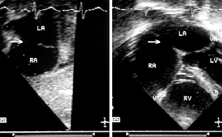
Figure 1 .
(Left) Short axis section of the atria, showing an oval fossa defect suitable for transcatheter closure. (Right) Four chamber section from the same patient, revealing an adequate rim of septum between the defect and the atrioventricular valves. A, left atrium; RA, right atrium; arrow, atrial septal defect; LV, left ventricle; RV, right ventricle; S, septum.
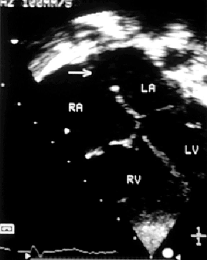
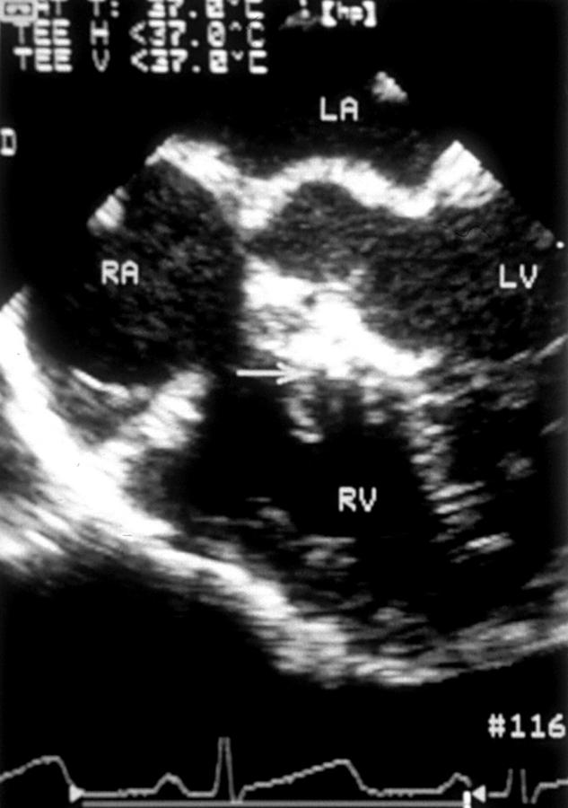
Figure 2 .
Four chamber section from an oval fossa atrial septal defect, in which there is a deficiency of infolding with an incomplete postero-superior rim. The arrow identifies the defect. LA, left atrium; RA, right atrium; LV, left ventricle; RV, right ventricle.
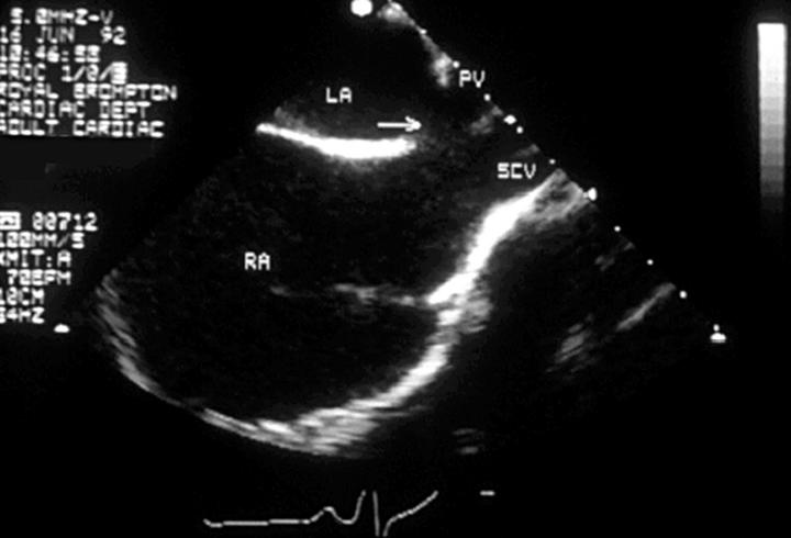
Figure 3 .
Transoesophageal echocardiogram in the vertical plane from a patient with a sinus venosus atrial septal defect, in which the superior caval vein (SCV) and right upper pulmonary vein (PV) override the atrial septum which divides the left atrium (LA) from right atrium (RA).
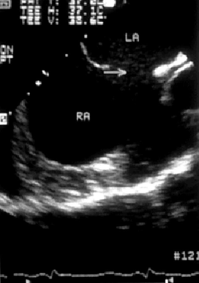
Figure 4 .
Transoesophageal echocardiogram in which an oval fossa atrial septal defect is indicated by an arrow. LA, left atrium, RA, right atrium.
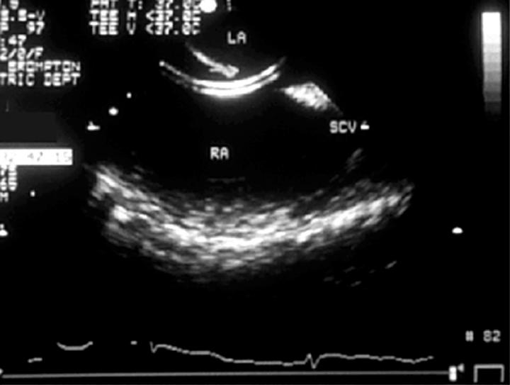
Figure 5 .
Transoesophageal echocardiogram identifying a catheter passing through a moderately large oval fossa atrial septal defect. LA, left atrium, RA, right atrium; SCV, superior caval vein.
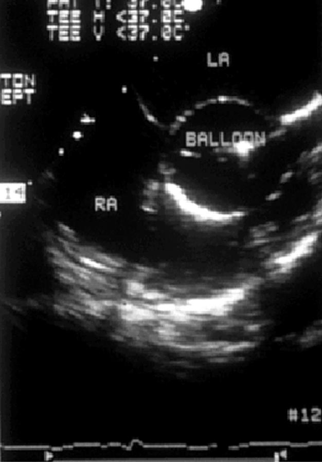
Figure 6 .
Transoesophageal echocardiogram recorded during balloon sizing of an oval fossa atrial septal defect. LA, left atrium, RA, right atrium.
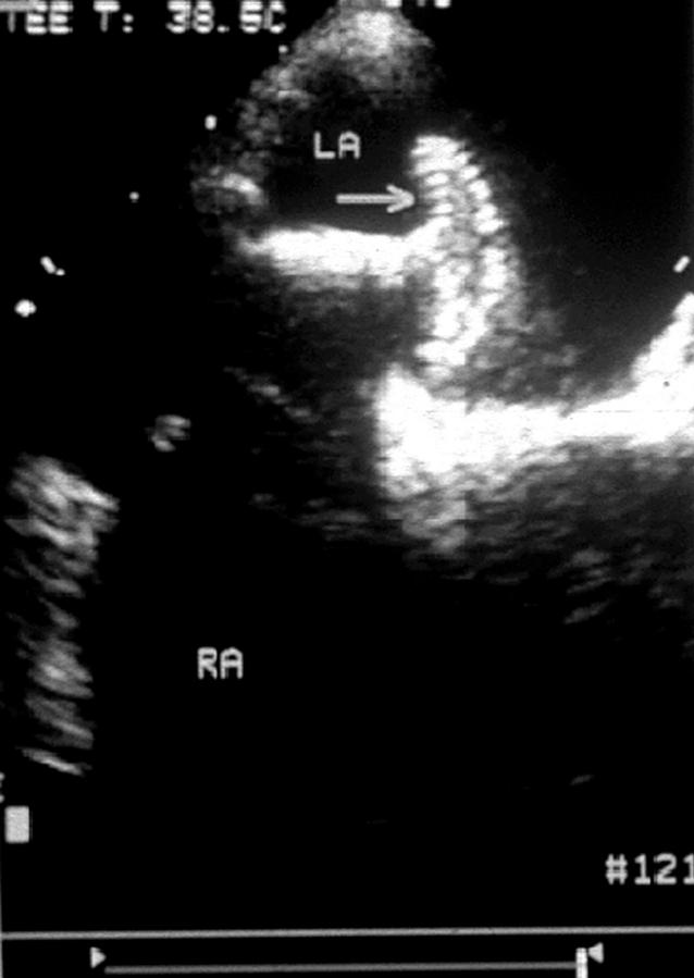
Figure 7 .
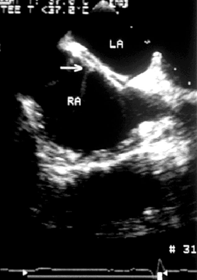
Transoesophageal echocardiogram recorded during deployment of a closure device illustrating the left atrial disc opened in the left atrium (arrow). LA, left atrium, RA, right atrium.
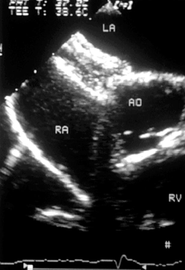
Figure 8 .
Transoesophageal echocardiogram recorded during device closure of an atrial septal defect with an Amplatzer occluder, showing left atrial and right atrial discs opened in their respective atria. AO, aortic root; LA, left atrium, RA, right atrium; RV, right ventricle.
Figure 9 .
Transoesophageal echocardiogram recorded during closure of an oval fossa defect with a Cardioseal device (arrow). LA, left atrium; RA, right atrium; LV, left ventricle; RV, right ventricle.
Figure 10 .
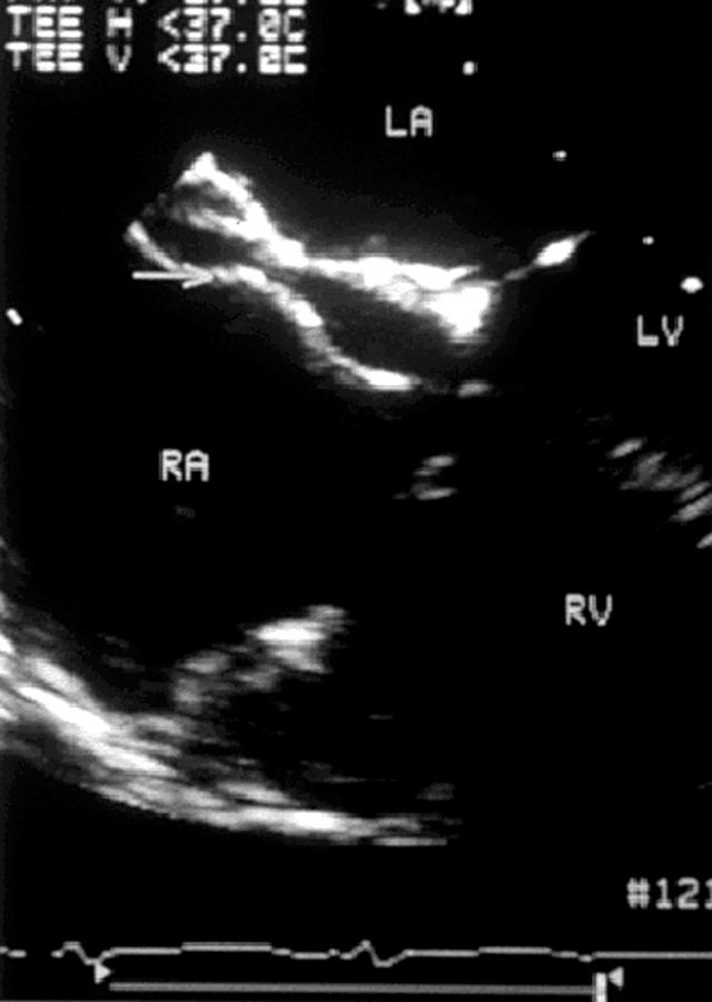
Transoesophageal echocardiogram recorded during closure of an oval fossa atrial septal defect with an Angel Wings device. LA, left atrium; RA, right atrium.
Figure 11 .
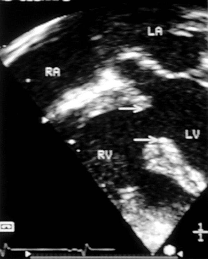
Transoesophageal horizontal section of a large muscular ventricular septal defect, whose borders are identified by the arrows. LA, left atrium; RA, right atrium; LV, left ventricle; RV, right ventricle.
Figure 12 .
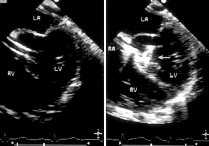
(Left) Transoesophageal echocardiogram recorded during device closure of a perimembranous ventricular septal defect, in which the sheath can be seen crossing the defect from right ventricle (RV) to left ventricle (LV). (Right) Horizontal plane transoesophageal echocardiogram recorded during device closure of a perimembranous ventricular septal defect, in which the left ventricular disc (arrow) can be seen to open within the left ventricle. LA, left atrium; RA, right atrium.
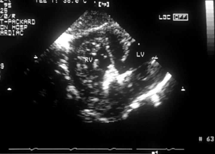
Figure 13 .
Horizontal transoesophageal echocardiogram following device closure of a perimembranous ventricular septal defect. An arrow identifies the position of the closure device within the ventricular septal defect.
Figure 14 .
Transgastric short axis echocardiographic section following transcatheter closure of a muscular ventricular septal defect, showing the closure device within the muscular defect. The right ventricular disc is well seen within the right ventricle (RV), although the left ventricular disc within the left ventricle (LV) is imaged less well.
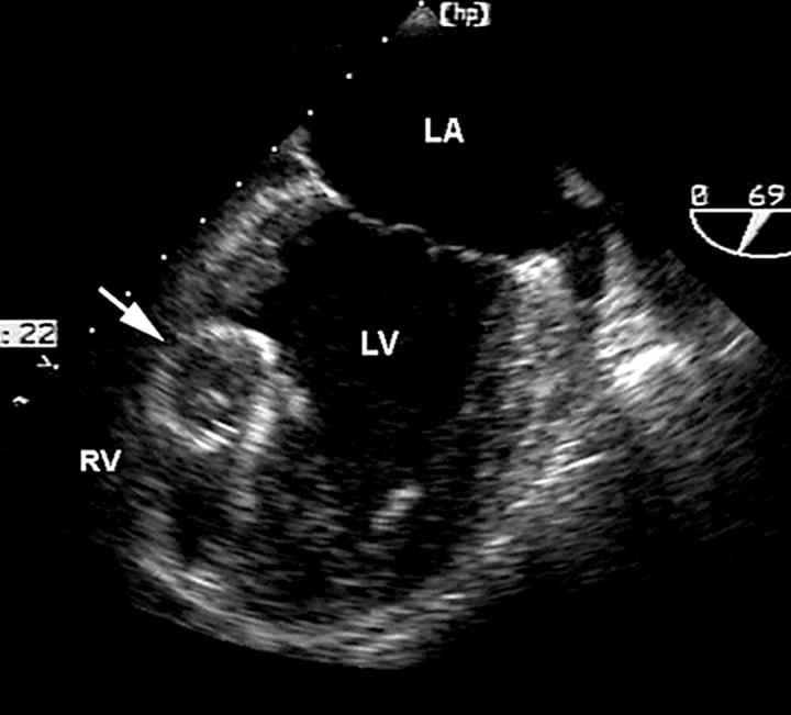
Figure 15 .
Transoesophageal echocardiogram recorded immediately following device closure of an ischaemic muscular ventricular septal defect in a patient presenting with cardiogenic shock. An arrow identifies the Amplatzer closure device. LA, left atrium; LV, left ventricle; RV, right ventricle.
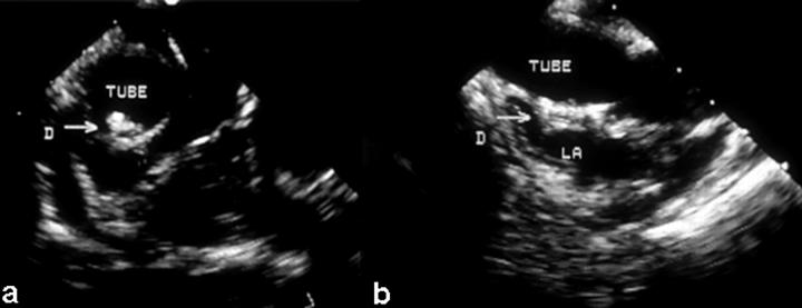
Figure 16 .
Transoesophageal echocardiograms immediately following transcatheter closure of a fenestrated lateral tunnel (tube) in a patient who had undergone a previous total caval pulmonary connection. The closure device (D, arrow) is seen in (a) horizontal and (b) vertical sections. LA, left atrium.
Selected References
These references are in PubMed. This may not be the complete list of references from this article.
- Dall'Agata A., McGhie J., Taams M. A., Cromme-Dijkhuis A. H., Spitaels S. E., Breburda C. S., Roelandt J. R., Bogers A. J. Secundum atrial septal defect is a dynamic three-dimensional entity. Am Heart J. 1999 Jun;137(6):1075–1081. doi: 10.1016/s0002-8703(99)70365-0. [DOI] [PubMed] [Google Scholar]
- Ewert P., Berger F., Vogel M., Dähnert I., Alexi-Meshkishvili V., Lange P. E. Morphology of perforated atrial septal aneurysm suitable for closure by transcatheter device placement. Heart. 2000 Sep;84(3):327–331. doi: 10.1136/heart.84.3.327. [DOI] [PMC free article] [PubMed] [Google Scholar]
- Ferguson J. J. American College of Cardiology 45th Annual Scientific Session, Orlando, Florida, March 24 to 27, 1996. Circulation. 1996 Jul 1;94(1):1–5. doi: 10.1161/01.cir.94.1.1. [DOI] [PubMed] [Google Scholar]
- Hijazi Z., Wang Z., Cao Q., Koenig P., Waight D., Lang R. Transcatheter closure of atrial septal defects and patent foramen ovale under intracardiac echocardiographic guidance: feasibility and comparison with transesophageal echocardiography. Catheter Cardiovasc Interv. 2001 Feb;52(2):194–199. doi: 10.1002/1522-726x(200102)52:2<194::aid-ccd1046>3.0.co;2-4. [DOI] [PubMed] [Google Scholar]
- Jan S. L., Hwang B., Lee P. C., Fu Y. C., Chiu P. S., Chi C. S. Intracardiac ultrasound assessment of atrial septal defect: comparison with transthoracic echocardiographic, angiocardiographic, and balloon-sizing measurements. Cardiovasc Intervent Radiol. 2001 Mar-Apr;24(2):84–89. doi: 10.1007/s002700000397. [DOI] [PubMed] [Google Scholar]
- Laussen P. C., Hansen D. D., Perry S. B., Fox M. L., Javorski J. J., Burrows F. A., Lock J. E., Hickey P. R. Transcatheter closure of ventricular septal defects: hemodynamic instability and anesthetic management. Anesth Analg. 1995 Jun;80(6):1076–1082. doi: 10.1097/00000539-199506000-00002. [DOI] [PubMed] [Google Scholar]
- Lock J. E., Block P. C., McKay R. G., Baim D. S., Keane J. F. Transcatheter closure of ventricular septal defects. Circulation. 1988 Aug;78(2):361–368. doi: 10.1161/01.cir.78.2.361. [DOI] [PubMed] [Google Scholar]
- Lock J. E., Rome J. J., Davis R., Van Praagh S., Perry S. B., Van Praagh R., Keane J. F. Transcatheter closure of atrial septal defects. Experimental studies. Circulation. 1989 May;79(5):1091–1099. doi: 10.1161/01.cir.79.5.1091. [DOI] [PubMed] [Google Scholar]
- Magni G., Hijazi Z. M., Pandian N. G., Delabays A., Sugeng L., Laskari C., Marx G. R. Two- and three-dimensional transesophageal echocardiography in patient selection and assessment of atrial septal defect closure by the new DAS-Angel Wings device: initial clinical experience. Circulation. 1997 Sep 16;96(6):1722–1728. doi: 10.1161/01.cir.96.6.1722. [DOI] [PubMed] [Google Scholar]
- Marx G. R., Fulton D. R., Pandian N. G., Vogel M., Cao Q. L., Ludomirsky A., Delabays A., Sugeng L., Klas B. Delineation of site, relative size and dynamic geometry of atrial septal defects by real-time three-dimensional echocardiography. J Am Coll Cardiol. 1995 Feb;25(2):482–490. doi: 10.1016/0735-1097(94)00372-w. [DOI] [PubMed] [Google Scholar]
- O'Dell S. D., Humphries S. E., Day I. N. Rapid methods for population-scale analysis for gene polymorphisms: the ACE gene as an example. Br Heart J. 1995 Apr;73(4):368–371. doi: 10.1136/hrt.73.4.368. [DOI] [PMC free article] [PubMed] [Google Scholar]
- Pedra C. A., Pihkala J., Lee K. J., Boutin C., Nykanen D. G., McLaughlin P. R., Harrison D. A., Freedom R. M., Benson L. Transcatheter closure of atrial septal defects using the Cardio-Seal implant. Heart. 2000 Sep;84(3):320–326. doi: 10.1136/heart.84.3.320. [DOI] [PMC free article] [PubMed] [Google Scholar]
- Perry S. B., Lang P., Keane J. F., Jonas R. A., Sanders S. P., Lock J. E. Creation and maintenance of an adequate interatrial communication in left atrioventricular valve atresia or stenosis. Am J Cardiol. 1986 Sep 15;58(7):622–626. doi: 10.1016/0002-9149(86)90288-2. [DOI] [PubMed] [Google Scholar]
- Pesonen E., Thilen U., Sandström S., Arheden H., Koul B., Olsson S. E., Wilson R. F., Toher C., Bank A., Bass J. Transcatheter closure of post-infarction ventricular septal defect with the Amplatzer Septal Occluder device. Scand Cardiovasc J. 2000 Aug;34(4):446–448. doi: 10.1080/14017430050196315. [DOI] [PubMed] [Google Scholar]
- Redington A. N., Rigby M. L. Transcatheter closure of interatrial communications with a modified umbrella device. Br Heart J. 1994 Oct;72(4):372–377. doi: 10.1136/hrt.72.4.372. [DOI] [PMC free article] [PubMed] [Google Scholar]
- Rickers C., Hamm C., Stern H., Hofmann T., Franzen O., Schräder R., Sievert H., Schranz D., Michel-Behnke I., Vogt J. Percutaneous closure of secundum atrial septal defect with a new self centering device ("angel wings"). Heart. 1998 Nov;80(5):517–521. doi: 10.1136/hrt.80.5.517. [DOI] [PMC free article] [PubMed] [Google Scholar]
- Sideris E. B., Walsh K. P., Haddad J. L., Chen C. R., Ren S. G., Kulkarni H. Occlusion of congenital ventricular septal defects by the buttoned device. "Buttoned device" Clinical Trials International Register. Heart. 1997 Mar;77(3):276–279. doi: 10.1136/hrt.77.3.276. [DOI] [PMC free article] [PubMed] [Google Scholar]
- Sigwart U. Alcohol ablation for mid-ventricular obstruction: straying from the path of righteousness? Eur J Echocardiogr. 2000 Dec;1(4):227–228. doi: 10.1053/euje.2000.0047. [DOI] [PubMed] [Google Scholar]
- Spevak P. J., Bass J. L., Ben-Shachar G., Hesslein P., Keane J. F., Perry S., Pyles L., Lock J. E. Balloon angioplasty for congenital mitral stenosis. Am J Cardiol. 1990 Aug 15;66(4):472–476. doi: 10.1016/0002-9149(90)90708-9. [DOI] [PubMed] [Google Scholar]
- Thanopoulos B. D., Tsaousis G. S., Konstadopoulou G. N., Zarayelyan A. G. Transcatheter closure of muscular ventricular septal defects with the amplatzer ventricular septal defect occluder: initial clinical applications in children. J Am Coll Cardiol. 1999 Apr;33(5):1395–1399. doi: 10.1016/s0735-1097(99)00011-x. [DOI] [PubMed] [Google Scholar]


