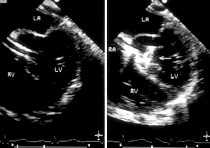
Figure 12 .
(Left) Transoesophageal echocardiogram recorded during device closure of a perimembranous ventricular septal defect, in which the sheath can be seen crossing the defect from right ventricle (RV) to left ventricle (LV). (Right) Horizontal plane transoesophageal echocardiogram recorded during device closure of a perimembranous ventricular septal defect, in which the left ventricular disc (arrow) can be seen to open within the left ventricle. LA, left atrium; RA, right atrium.

