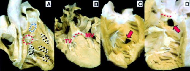
Figure 15 .
(A) The locations of perimembranous (red circles), muscular (black circles), and doubly committed (blue circles) subtypes of ventricular septal defects are superimposed on this right ventricular view. (B) This four chamber cut through a perimembranous inlet ventricular septal defect shows the fibrous continuity (red dotted line) between tricuspid (TV) and mitral (MV) valves forming the roof of the defect. The normal offset between the valves is lost. (C) The muscular defect (arrow) has complete muscular borders. (D) The doubly committed and juxta-arterial defect (arrow) is roofed by fibrous continuity (red dotted line) between the aortic and pulmonary valves.

