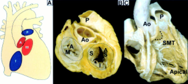
Figure 3 .
(A) The pulmonary (P), aortic (A), mitral (M), and tricuspid (T) valves are offset from one another. (B) When viewed from the base, the aortic valve(Ao) is centrally located and tucked into the waist of the figure-of-eight arrangement formed by mitral and tricuspid valves. The aortic, or "anterior", leaflet (A) of the mitral valve occupies a shorter circumference than the mural (M) leaflet. The three leaflets of the tricuspid valve are designated septal (S), anterosuperior (AS), and inferior (I). (C)The right heart valves are separated by the ventriculo-infundibular fold (dotted line) which lies between the limbs of the septomarginal trabeculation (SMT). Note the free standing muscular sleeve underneath the pulmonary valve that is the outlet portion of the right ventricle. The inlet portion contains the tricuspid valvar apparatus and the apical portion is coarsely trabeculated. Arrow = moderator band.

