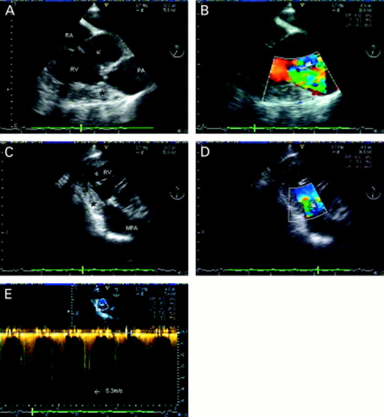
Figure 9 .

Mid cavity right ventricular obstruction. (A) Short axis view just below the aortic valve. A large perimembraneous ventricular septal defect is seen (arrow) between the left ventricular outflow tract and inlet part of the right ventricle. There is severe right ventricular hypertrophy and a critical obstruction in mid-cavity (*). The pulmonary valve leaflets cannot be seen; the level of the annulus is indicated (PA). In a similar view with colour flow mapping (B), flow convergence and turbulence are seen within the right ventricular cavity in systole, at the site of the obstruction (arrow). In the transgastric view, a long axis view of the right ventricle can be obtained, which shows the relation of the obstruction (*) to the main pulmonary artery (C) and allows good alignment for colour flow (D) and continuous wave Doppler (E). A peak gradient of 118 mm Hg was measured. RA, right atrium; RV, right ventricle; MPA, main pulmonary artery; PA, pulmonary annulus.
