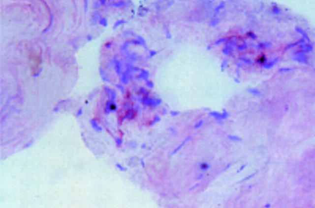
Figure 1 .
Immunohistological examination of a sacroiliac biopsy specimen obtained by computed tomography guided biopsy of a 23 year old patient with ankylosing spondylitis, four years disease duration, with inflammatory back pain grade 6 on a visual analogue scale (0-10). The red staining indicates a mononuclear cell positive for TNFα (APAAP staining technique).

