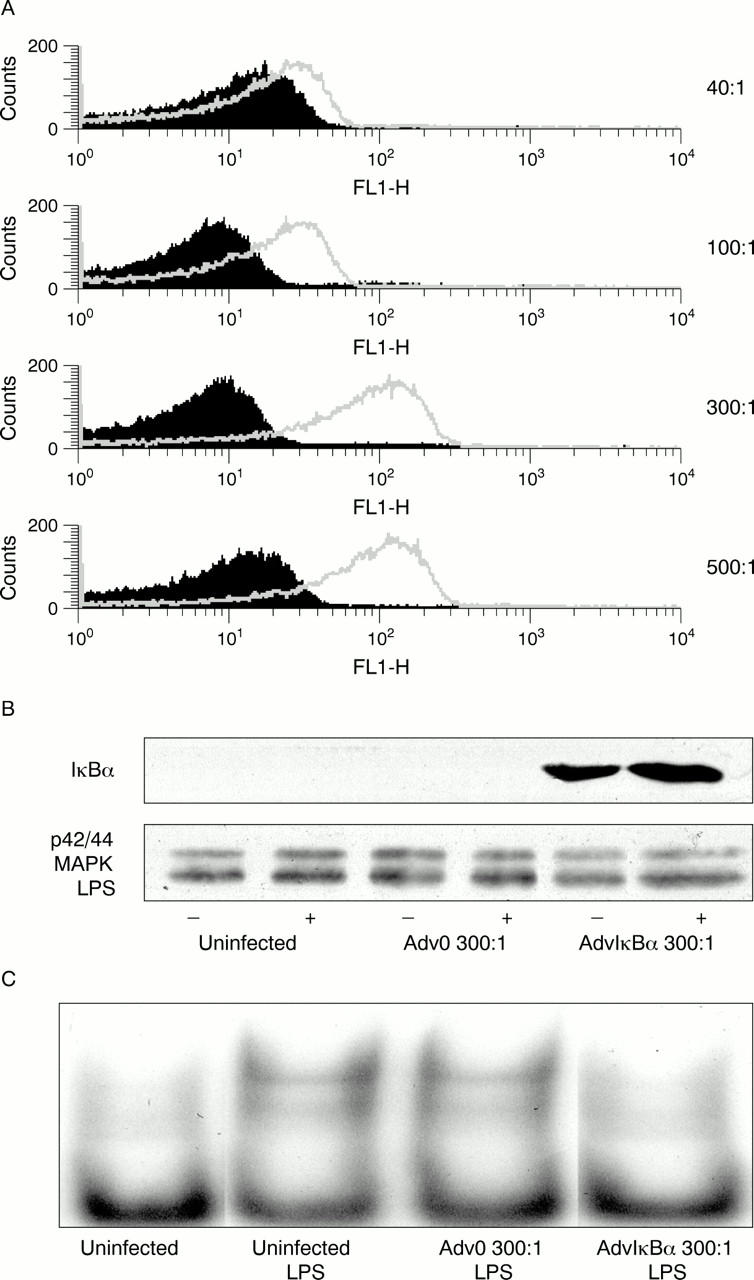
Figure 1 .

(A) Mature dendritic cells were generated by five days' culture in 50 ng/ml granulocyte macrophage colony stimulating factor and 10 ng/ml interleukin 4 (IL4) and a further three days in monocyte conditioned medium. Then they were plated on a 96 well, flat bottom plate at a density of 2×10⁵ cells/well and left either uninfected with a multiplicity of infection ranging from 40 to 500 of an adenovirus without insert (Adv0) or an adenovirus overexpressing β-Gal expression. Fluorescein-di-(β-D)-galactopyranoside (Sigma) was used as a substrate of β-galactosidase and cell fluorescence was analysed by FACS analysis. In (B) and (C), 10×106 cells per condition were left either uninfected or infected with Adv0 or AdvIκBα at a multiplicity of infection of 300. Two days after infection, cells were left unstimulated or stimulated with lipopolysaccharide (LPS; 50 ng/ml) for 60 minutes. Cytosolic and nuclear extracts were then prepared and examined for IκBα or p42/44MAPK (loading control) expression by sodium dodecyl sulphate-polyacrylamide gel electrophoresis (B), or for NF-κB DNA binding activity by electrophoretic mobility shift assay (C).
