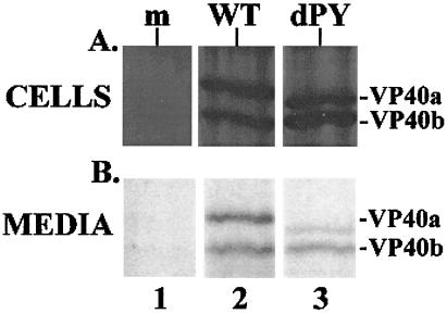
Figure 6.
VP40 budding assay. (A) Radiolabeled cell extracts from COS-7 cells that received no DNA (mock, lane 1), pVP40-WT (lane 2), and pVP40-dPY (lane 3) were immunoprecipitated and analyzed by SDS/PAGE. (B) Radiolabeled proteins released into the media covering cells transfected with no DNA (mock, lane 1), pVP40-WT (lane 2), and pVP40-dPY (lane 3) were immunoprecipitated as above. Immunoprecipitated proteins were quantitated using National Institutes of Health image Version 2 software.

