
Figure 3.
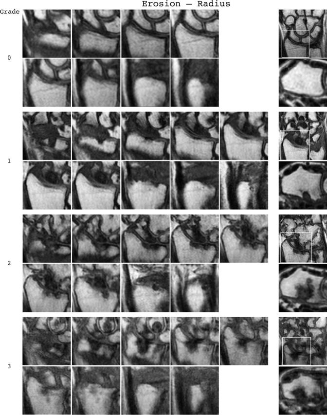

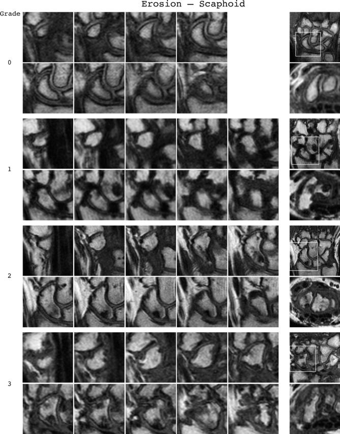

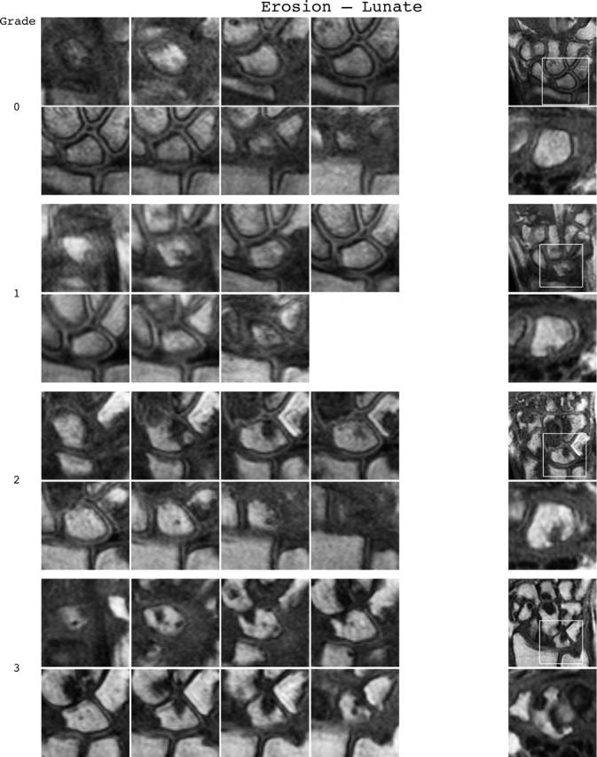

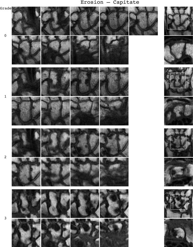

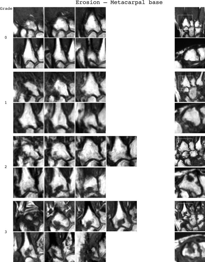


Bone erosion reference image sheets (pages i36–i45, total 10). Bone erosion in the radius, scaphoid, lunate, capitate and a metacarpal base is illustrated on two single-page sheets each. Grades 0–3 supplemented with examples of three higher grades are provided. Bone erosion is graded by assessing percentage volume (1–10, by 10% volume increments) of the assessed bone volume as described in the OMERACT RAMRIS (see table 1, reference 17). The "assessed bone volume" is defined as described above. It should be emphasised that all coronal slices covering the bone should be assessed to estimate the percentage of the total volume occupied by the erosion. The atlas reference images can be used for guidance and calibration. Each bone of the wrist should be scored separately. A total score (range 0–150) can be calculated. The drawing above explains the types and positions of images presented. The varying number of coronal slices needed to cover the bone reflects varying bone sizes and varying slice thickness (2–3 mm).