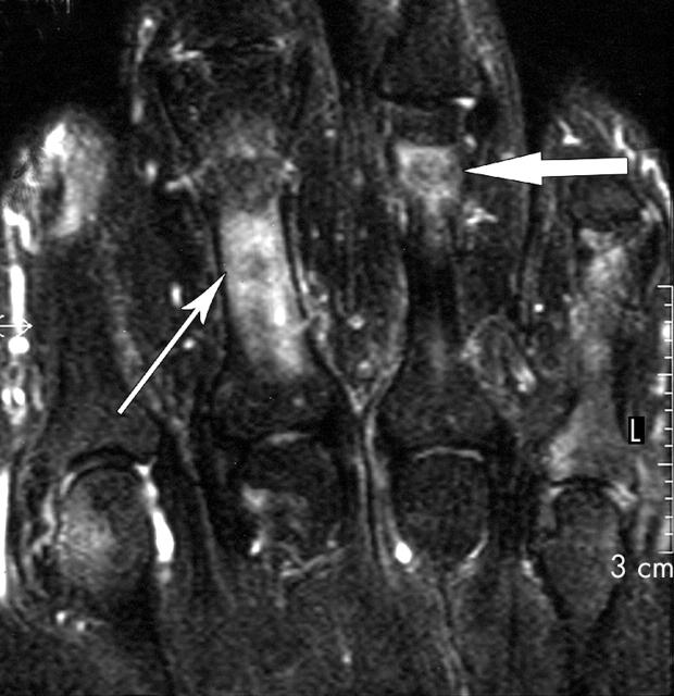
Figure 16.
Short tau inversion recovery (STIR) coronal image of the fingers of a patient with rheumatoid arthritis covering the second to fifth metacarpophalangeal and proximal interphalangeal joints. Bright signal over the proximal phalanx of the third finger (thin arrow) resembles bone oedema but is actually due to inflammation within the flexor tendon sheath (tenosynovitis). True bone oedema is seen distally at the fourth proximal phalanx (wide arrow).

