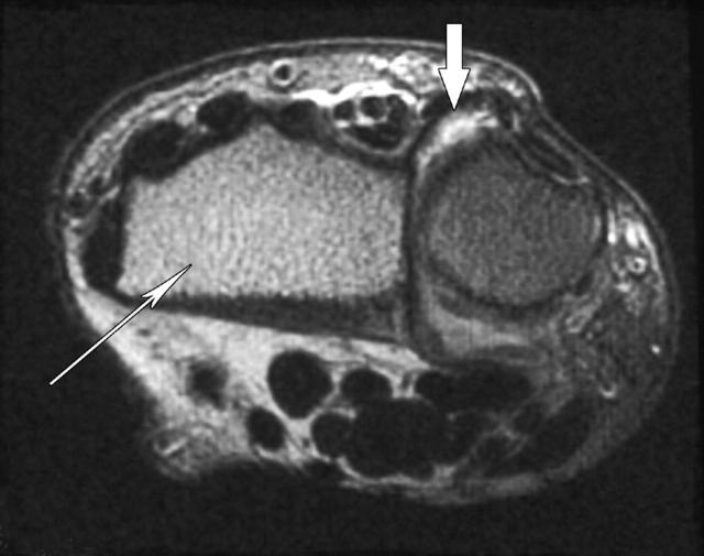
Figure 4.
T2 weighted axial image of the distal radius and ulna. Fat saturation is complete for the ulna but not for the radius, where "shine through" of marrow fat is apparent (thin arrow). This appearance mimics bone oedema. Fat in subcutaneous tissues is also bright in the left half of the picture but not in right half where bright signal in the distal radioulnar joint indicates synovitis (wide arrow).

