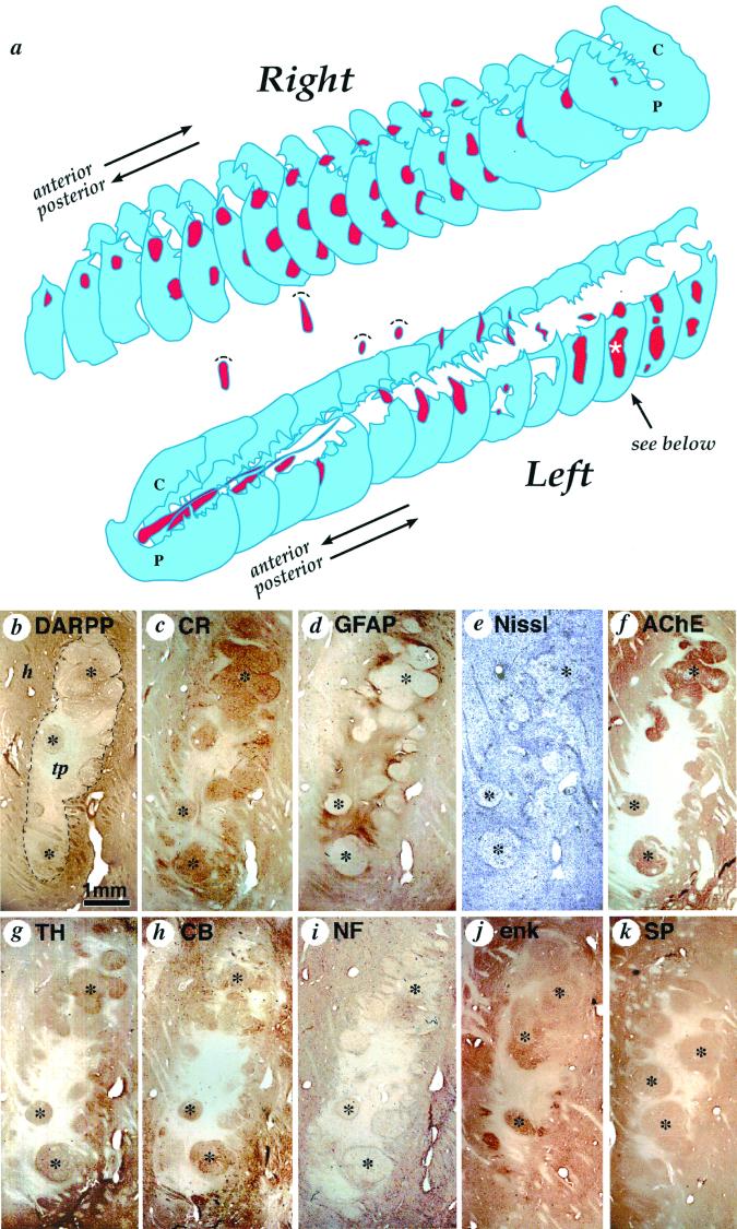
Figure 1.
(a) Three-dimensional schematic reconstruction of the patient's caudate nucleus and putamen illustrating the sites and relative sizes of bilateral human fetal cell implants as they appeared 18 months after implantation. Caudate nucleus and putamen cross-sections are shown in blue, and transplant tissue is shown in red. Two identified cell implants in the left putamen and three identified transplants in the right putamen were columnar in shape and oriented horizontally following bilateral frontal tracks. The rostral-most left hemisphere tract, intended for the caudate nucleus, was confined to the internal capsule. Volume of this implant was comparatively reduced and more cell dense than any other graft. (b–k) Serial sections through the most posterior graft in the left putamen (indicated by a white asterisk in a), stained with different neurohistological markers to reveal transplant architecture and immunohistochemically stained with antibodies characteristic of striatal neural cell phenotypes: (b) dopamine and cAMP-associated receptor phosphoprotein, (c) calretinin, (d) glial fibrillary acidic protein for astrocytes, (e) cresyl violet (Nissl) stain for general cell perikarya, (f) acetylcholinesterase staining for cholinergic cells and innervation (also see fig. 2a for choline acetyltransferase), (g) tyrosine hydroxylase staining for donor tissue axons, (h) calbindin, (i) 200-kDa human neurofilament (NF-200) for mature axons, (j) enkephalin, and (k) substance P. Cell implant cytoarchitecture (delineated by a dotted line in a) is characterized by two distinct tissue types: roughly spherical zones containing clusters of larger lower density neurons that are acetylcholinesterase-positive (f), glial fibrillary acidic protein-negative (d), and lightly neurofilament-positive (i) (marked by black stars); surrounded by regions of relatively high cell density that are acetylcholinesterase-negative, glial fibrillary acidic protein-negative -positive, and neurofilament-negative. The remaining photomicrographs identify overlapping classes of striatal phenotypes, whereas tyrosine hydroxylase stains host axons that specifically innervate striatal but not nonstriatal tissue. h, host; tp, transplant. (Scale bar = 1 mm.)

