
Figure 1.
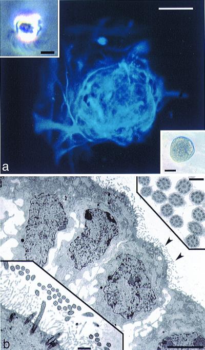
Generation of spherical clones from single, ciliated ependymal cells. (a) GFAP immunofluorescence (blue, AMCA) of a clone (phase microscopy of such a clone is shown in the lower right Inset), derived from a single ciliated ependymal cell (Inset, upper left; notice cilia protruding from the base of the cell). Scale bar in a and lower right Inset = 50 μm; scale bar in upper left Inset = 5 μm. (b) TEM of a clone derived from a single ciliated ependymal cell. Note cilia on the surfaces of cells at the edge of this clone (arrowheads), shown at higher magnification in the lower left Inset. The upper right Inset shows cross-sectioned cilia displaying the characteristic 9 + 2 arrangement of microtubules. Scale bar in b = 5 μm, 0.5 μm in the lower left Inset, and 0.2 μm in the upper right Inset.