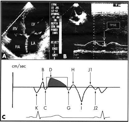
Figure 1.
(A) A four chamber view in a patient: the M mode cursor or sample volume is located at the junction of the right ventricular free wall and the tricuspid annular plane. (B) Schematic illustration of atrioventricular plane motion by M mode. (C) Schematic illustration of atrioventricular plane motion by pulsed Doppler. Adapted from reference 9. LA, left atrium; LV, left ventricle; RA, right atrium; RV, right ventricle; TPSE, tricuspid plane systolic excursion.

