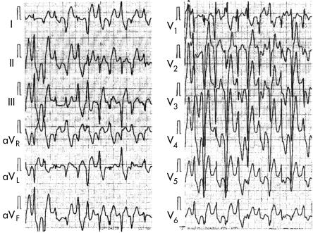
Figure 3.
Bidirectional ventricular tachycardia. This is the treadmill exercise ECG of a 12 year old boy who had syncope. Note the typical bidirectional ventricular tachycardia recorded in leads V5 and aVR; however, leads aVL and V1 showed polymorphic ventricular tachycardia. See text for further discussion.

