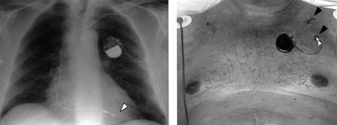
Figure 1.
(Left) Chest radiograph showing the position of the bipolar ventricular pacing lead positioned in the right ventricular apex using an active fixation screw (arrow). (Right) The lead was tunnelled subcut- aneously from the point of vascular access (top arrow) to an exit point approximately 10 cm away. The lead was secured to skin using the suture sleeve (bottom arrow) and was connected to an external permanent pacemaker pulse generator. Both lead and generator were covered with an adhesive dressing.

