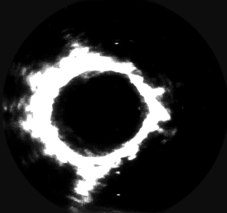
Figure 1.

Cross sectional IVUS image from a patient with primary pulmonary hypertension. Note the intima + media thickening (soft echoes) between the arterial lumen and the outer echobright layer (adventitia). Wall thickening is eccentric, with more prominent changes between 5 and 12 o’clock. Distance between two white points is 1 mm. The circular artefact within the lumen is generated by the ultrasound catheter.
