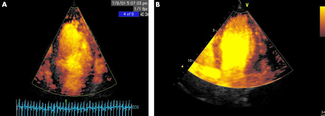
Figure 3.
Myocardial contrast echocardiography utilising intermittent power Doppler imaging triggered at end systole, after injection of a 0.5 ml bolus of Optison. Apical 4 chamber views. (A) Patient with recent anteroseptal myocardial infarction treated with thrombolysis but no clinical evidence of reperfusion. Absence of perfusion in septum and apex, compared to apical lateral segment, is readily appreciated. There is some dropout in the basal lateral segment caused by attenuation from apical contrast. (B) Patient with anteroseptal myocardial infarction treated successfully with thrombolysis. Normal contrast uptake in septum with minor apical defect only.

