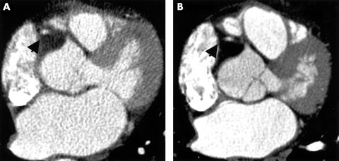
Figure 1.
Electron beam computed tomographic coronary angiography (EBCTA; panel A) and multislice computed tomographic coronary angiography (MSCTA; panel B) in the same 62 year old patient. On this axial image at the level of the proximal right coronary artery (RCA) the differences in contrast to noise ratio (CNR) and spatial resolution between the two methods are visualised. The arrow indicates the right coronary artery.

