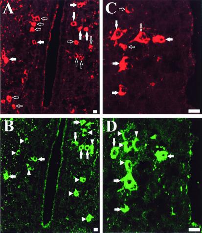
Figure 1.
Confocal laser scanning microscope photomicrographs comparing the distribution of 3β-HSD- and GABAA receptor α3 subunit-like immunoreactivity in the frog hypothalamus. (A and B) Adjacent frontal sections through the anterior preoptic area are labeled with the antiserum against 3β-HSD (A) or the polyclonal antibodies against the α3 subunit (B). (C and D) Adjacent frontal sections through the posterior tuberculum labeled with the antiserum against 3β-HSD (C) or the polyclonal antibodies against the α3 subunit (D). Open arrows, neurons expressing only the 3β-HSD-like immunoreactivity; arrowheads, neurons expressing only the GABAA receptor α3 subunit-like immunoreactivity; filled arrows, neurons expressing both 3β-HSD- and GABAA receptor α3 subunit-like immunoreactivities. (Bars = 10 μm.)

